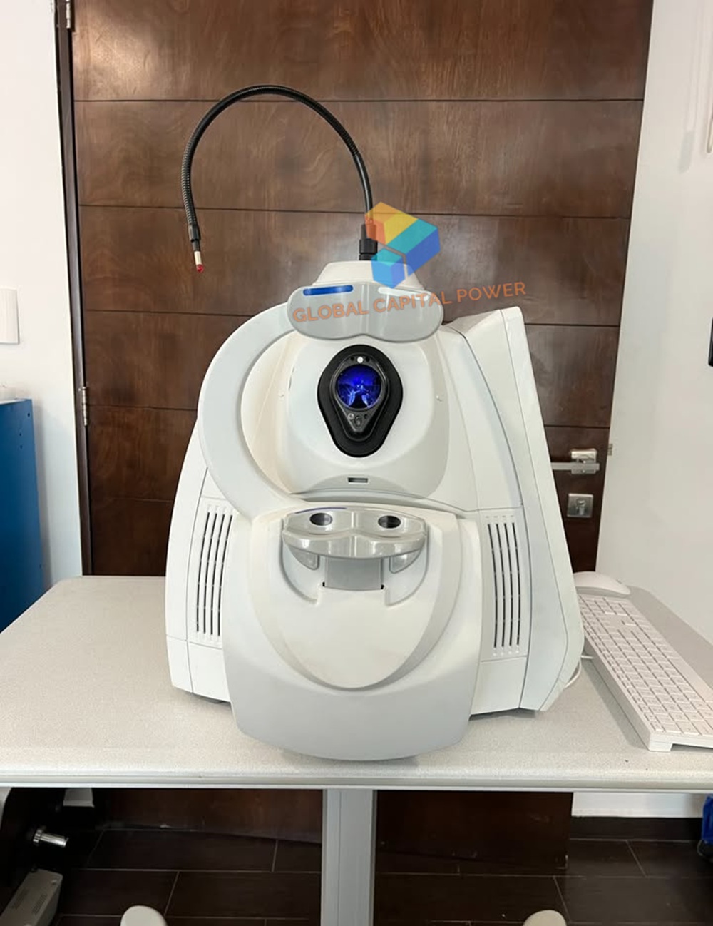
Carl Zeiss Cirrus 5000 OCT Imaging System
Original price was: $12,244.$6,122Current price is: $6,122. & Free Shipping
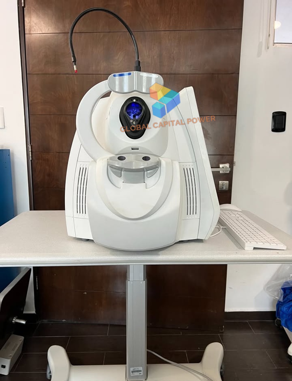
For Sale – Carl Zeiss Cirrus 5000 OCT Imaging System
High resolution visualization capabilities and sophisticated clinical applications. These include Advanced RPE analysis to track retinal pigment epithelial integrity and Ganglion Cell Analysis to assess glaucomatous loss in the macula. FastTrac retinal tracking helps prevent eye motion artifacts.
Free shipping on orders over $50!
- Satisfaction Guaranteed
- No Hassle Refunds
- Secure Payments
Description
Carl Zeiss Cirrus 5000 OCT Imaging System
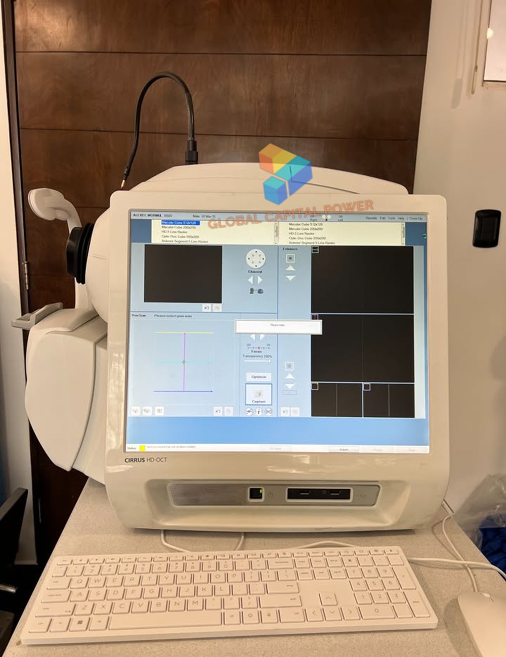
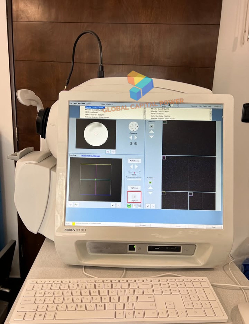
Carl Zeiss Cirrus 5000 OCT Imaging System features state-of-the-art hardware and software based on a fast 64-bit Windows 7 computer, a significantly accelerated OCT camera system, a much larger 19-inch monitor, and a wireless mouse and mouse Keyboard. This results in faster processing with shorter times for the patient and ophthalmologist. The OCT scan speed is between 27,000 and 68,000 per second.
Features
PanoMap Wide-Field Analysis
Integrated Insights
Backward Compatibility
Full Anterior Chamber Imaging
HD Cornea Scan
Pachymetry Map
Wide Angle-to-Angle Scan and HD Angle Scan
Smart HD 1 Line Scan
VRI en Face Preset Display
IS/OS-Ellipsoid en Face Preset Display
Specifications
OCT Imaging
Methodology: Spectral domain OCT
Optical source: Superluminescent diode (SLD), 840 nm
Scan speed: 27K- 68K A-scans per second
A-scan: 2.0 mm (in tissue), 1024
Axial resolution: 5 μm (in tissue)
Transverse resolution: 15 μm (in tissue)
Fundus Imaging
Methodology: Line scanning ophthalmoscope (LSO)
Live fundus image: During alignment and during OCT scan
Optical source: Superluminescent diode (SLD), 750 nm
Field of view: 36 degrees W x 30 degrees H
Frame rate: > 20 Hz
Transverse resolution: 25 μm (in tissue)
Iris Imaging
Methodology: CCD camera
Resolution: 1280 x 1024
Efficiency
Field of view
OCT Angiography
Anterior capability
Data migration
Empower your legacy data
The ZEISS CIRRUS platform protects legacy data, guided progression analysis and base lines which empower diagnostic decision-making. Technological advancements in speed, scan protocols and efficiencies found within the ZEISS CIRRUS enable you to provide patients with seamless continuity at every step of their care, when referring to legacy CIRRUS data.
Multi-modal OCT with full anterior chamber imaging and measurement capability
ZEISS CIRRUS is the first retinal OCT with full anterior chamber imaging and measurement capability. It can expand your ability to include comprehensive imaging and quantification of the anterior segment for refractive surgery planning and follow-up, corneal evaluation, and glaucoma assessment.
Include Purchase
Cirrus HD-OCT 5000 Unit
New Printer
Mouse
Keyboard
Warranty Table
Table
all peripherals
Related products
-
Sale!
-
Sale!
-
Sale!
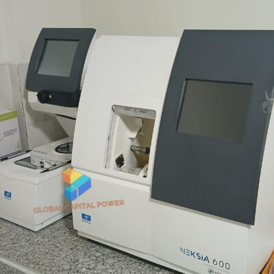
Essilor Neksia 600 Edging System
Original price was: $12,660.$6,330Current price is: $6,330. -
Sale!
iSee 9000 Dental Surgical Microscope
Original price was: $12,660.$6,330Current price is: $6,330.
Reviews
There are no reviews yet.