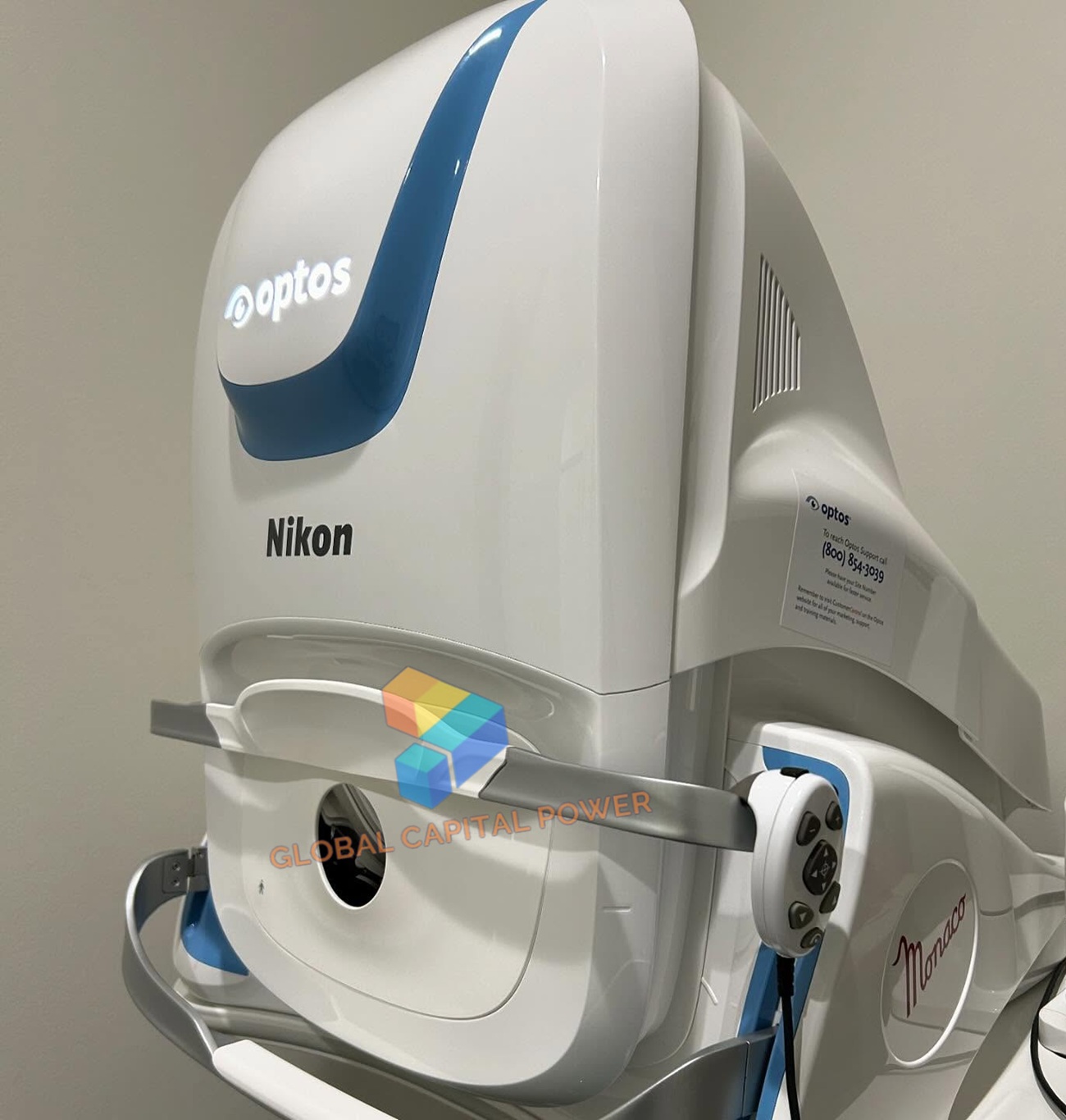

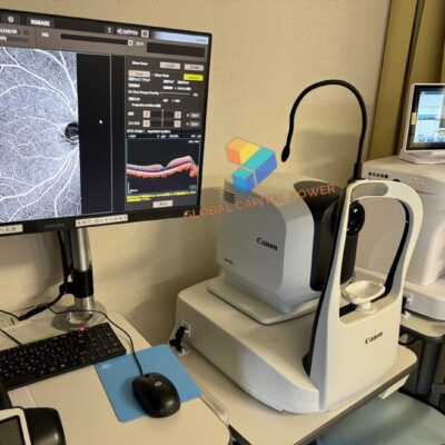
Nikon Optos Monaco Ultra WideField Retina Imaging
Original price was: $10,420.$5,210Current price is: $5,210. & Free Shipping
For Sale – Nikon Optos Monaco enables a rapid multi-modality capture featuring color, autofluorescence, and OCT scans, for both eyes, in as little as two minutes.
Free shipping on orders over $50!
- Satisfaction Guaranteed
- No Hassle Refunds
- Secure Payments
Description
Nikon Optos Monaco Ultra WideField Retina Imaging
Nikon Optos Monaco Ultra WideField Retina Imaging to perform OCT scanning. The accession of OCT”offers a broader collection of imaging tools within 1 machine,” the firm said in a press release,”enabling eye care practitioners to treat and see retinal pathology earlier and more efficiently.”
Features and Benefits
UWF with integrated OCT saves time, space and minimizes patient movement.
High resolution 200º single-capture optomap images improve pathology detection and management frommacula through the far periphery.
Non-mydriatic, cSlO imaging through most cataracts and small pupils (2 mm).
3-in-1 Color Depth ImagingTM provides important clinical data from the retinal surface through the choroid.
Green laser autofluorescence minimizes patient exposure to blue light and shows macula and optic nerve head detail.
Central pole OCT provides comprehensive multi-modal imaging.
optomap images and OCT scans are correlated to facilitate pathology examination.
Color, AF, and OCT images are shown in a single, comprehensive view.
Fast, comfortable image acquisition is easier on patients and improves clinic flow.
OptosAdvance Image Management facilitates image review and consultations and includes measurement and auto montage capabilities.
Image Modalities
optomap color and optomap plus (red and green laser):
Color composite view
Green laser view
Red laser view
optomap af (green laser): autofluorescence
Optical Coherence Tomography (OCT)
Resolution
optomap color: 20 μm
optomap plus, af : 14 μm
Exposure Time
Less than 0.4 seconds
OCT Scan Types
Line Scan
Raster Scan
Retina Topography Scan
Optic nerve Head (OnH) Topography Scan
Retinal nerve Fiber layer (RnFl) Scan
Wavelengths
Red laser: 635 nm
green laser: 532 nm (for af )
Tomographic Imaging
Signal Type: Optical scattering from tissue
Signal Source: Super luminescent Diode (SlD) 840 nm
Optical Power: laser safety Class-1 following IEC/en60825-1:2014
Typical Axial Resolution: <10 micron (in tissue) Digital on-screen <6 micron
Transverse Resolution: 20 micron (in tissue)
Scanners: Galavanometric with x, y mirrors
Scan Depth: Up to 2.5mm
OCT Scan Characteristics
Spectral Domain OCT
A-Scan rate up to 70k cycles/s
Active eye tracking
Automatic scan positioning
Footprint
Width: 550 mm/22 inches
Depth: 570 mm/23 inches
Height: 606 – 632 mm/24-25 inches
Related products
-
Sale!
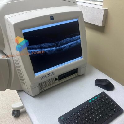
Carl Zeiss Cirrus 4000 OCT Imaging System
Original price was: $10,420.$5,210Current price is: $5,210. -
Sale!
-
-
Sale!
Reviews
There are no reviews yet.